
Comprehensive Eye Exams & So Much More

Our eye care center offers a full range of eye health services to clients of all ages, from children to older adults: routine eye exams and vision tests, eye disease diagnosis and management, immediate attention for eye emergencies, and complete eye health management. The optometrists in our practice provide eyewear prescriptions and offer corrective laser eye surgery co-management as well. You may call one of our three locations to set up an appointment at your convenience and learn more about some of our key services below.
Our Eye Care Services:
Eye Exams
Routine exams are important, regardless of your age or physical health. During a complete eye exam, your eye doctor will not only determine your prescription for eyeglasses or contact lenses, but will also check your eyes for common eye diseases, assess how your eyes work together as a team, and evaluate your eyes as an indicator of your overall health. The American Optometric Association (AOA) also recommends an annual eye exam for any adult who wears eyeglasses or contacts. If you don't normally need vision correction, you still need an eye exam every year. Doctors often recommend more frequent examinations for adults with diabetes, high blood pressure, and other disorders because many diseases can have an impact on vision and eye health.
Pediatric Eye Exams
Some experts estimate that approximately 5% to 10% of pre-schoolers and 25% of school-aged children have vision problems. According to the AOA, all children should have their eyes examined at 6 months of age, at age 3, and again at the start of school. Children without vision problems or risk factors for eye or vision problems should then continue to have their eyes examined annually throughout their school years.
The InfantSEE® program offers a one-time, comprehensive eye and vision assessment to babies in their first year of life, usually between the ages of 6 and 12 months, offering early detection of potential eye and vision problems at no cost regardless of income or ability to pay.
Eye Emergencies
We are always willing to help should you ever experience an eye emergency. Our offices provide emergency services for eye infections, eye injuries, and other eye urgencies. We accommodate many eye emergencies such as:
- Foreign materials stuck in the eyes
- Eye trauma
- Scratched eyes
- Sudden loss of vision in one or both eyes
- Flashes of light in the vision
- “Floaters” in the vision
- Red or painful eyes
- Uncomfortable, itchy, or irritated eyes
Studies have shown that an overwhelming number of emergency room visits could have been treated by an optometrist. It is not always necessary to go to an emergency room for eye emergencies. Optometrists are equipped to treat the majority of eye emergencies. Please call one of our offices and we will schedule you as soon as possible.
If it is during our office hours, please contact us immediately at one of our three locations for guidance. We’ll help you with the best treatment to prevent complications and promote long-lasting clear eyesight.
For an after hour ocular emergency, please call 937-492-9197 for further instructions. Use your best judgment on urgency as you may find the need to go to the nearest emergency room.
Eye Disease Management
Your eye doctor will also monitor any eye health-related issues discovered during your routine exam. We also provide eye health-related exams to manage longstanding conditions such as, but not limited to:
- Diabetic retinopathy
- Glaucoma
- Cataracts
- Macular degeneration
- Dry eye syndrome
- Corneal dystrophies
Our state of the art equipment allows us to examine the front surface of the eye and also digitally scan inside the eye to monitor for changes in your eye health and help the doctor determine a treatment plan if you are diagnosed with an eye disease. It is important to have an annual eye exam to continue monitoring your eyes for any health-related changes. Your eye doctor may have you return more frequently if you are diagnosed with an eye disease.
Our Diagnostic Equipment:
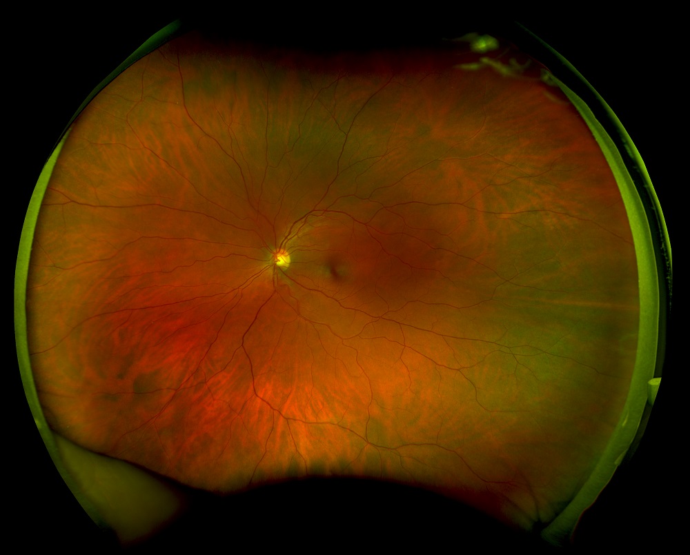
Optomap For many patients, having pupils dilated using eye drops can be a bother. But as an integral part of a truly comprehensive eye exam, those drops are highly recommended. Dilation gives your eye doctor the widest view of the internal structures at the back of the eye—the optic nerve, retina, even blood vessels.
That’s where Optomap technology comes in. Using low-power laser technology, your eyecare professional can take a wide, instantly-viewable and detailed digital scan of your retina (the area responsible for processing images). All in real time. And in no time. Without the use of pupil-dilating eye drops.

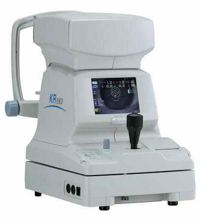
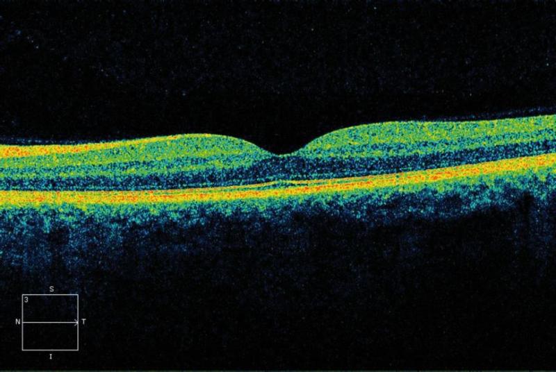
Optical Coherence Tomography (OCT) is a non-invasive diagnostic instrument used for imaging the retina. It is the technology for the future because it can enhance patient care. It has the ability to detect problems in the eye prior to any symptoms being present in the patient. With an OCT, doctors are able to see a cross section or 3D image of the retina and detect the early onset of a variety of eye conditions and eye diseases such as macular degeneration, glaucoma and diabetic retinopathy (the top three diseases known to cause blindness).
The OCT has become the standard of care for the assessment and treatment of most retinal diseases, it is similar to a CT scan which is used to image internal organs inside the body. The OCT uses an array of light to rapidly scan the eye. These scans are interpreted and the OCT then presents an image of the tissue layers within the retina. These layers can be differentiated and their thickness measured. By comparing the thickness of the layers measured by the OCT scan against the normal thickness of healthy retinal layers, eye doctors can determine which retinal disease or eye condition exists in the eye, even before the patient is aware of any problems.

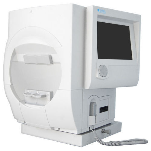
Visual Field testing is a method of measuring an individual’s central and peripheral (side) vision. Visual field testing is most frequently used to detect any signs of glaucoma damage to the optic nerve. In addition, visual field tests are useful for detection of central or peripheral retinal disease, eyelid conditions such as ptosis or drooping of the eyelid, optic nerve disease, and diseases affecting the perimeter.
To do the test, you sit and look inside a bowl-shaped instrument called a perimeter. While you stare at the center of the bowl, lights flash. You press a button each time you see a flash. A computer records the spot and at the end of the test, a printout shows if there are areas of your vision where you did not see the flashes of light. These are areas of vision loss. If you are diagnosed with a particular disorder or disease such as glaucoma, visual field testing will become a routine part of your treatment. After a baseline has been established, visual field testing is repeated every 6 to 12 months to monitor for change. Visual field tests play a critical role in helping the doctor follow your condition.

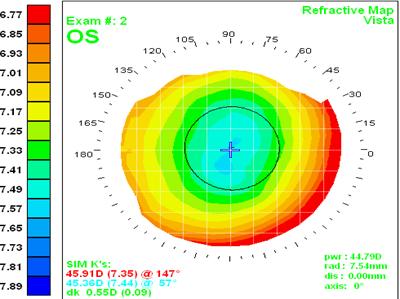
Corneal topography is a computer assisted diagnostic tool that creates a three-dimensional map of the surface curvature of the cornea. The cornea (the front window of the eye) is responsible for about 70 percent of the eye’s focusing power. An eye with normal vision has an evenly rounded cornea, but if the cornea is too flat, too steep, or unevenly curved, less than perfect vision results. The greatest advantage of corneal topography is its ability to detect irregular conditions invisible to most conventional testing.
Corneal topography produces a detailed, visual description of the shape and power of the cornea. This type of analysis provides your doctor with very fine details regarding the condition of the corneal surface. These details are used to diagnose, monitor, and treat various eye conditions. They are also used in fitting specialty contact lenses.